Heavily calcified mesial lingual canal on #37
This was a challenging case.
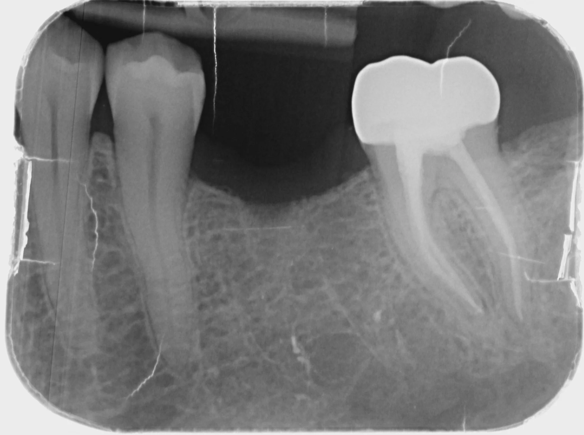
A diagnosis of pulp necrosis with apical periodontitis was made on tooth 37. Preoperative periapical radiograph is fairly inconspicuous with pulp canal spaces fairly visible.

Preoperative radiograph of #37.
However the CBCT slices show heavy calcification of mesial buccal canal in coronal 1/3. The orifice to ML canal is likely to be buried underneath 3-4mm of tertiary dentine.
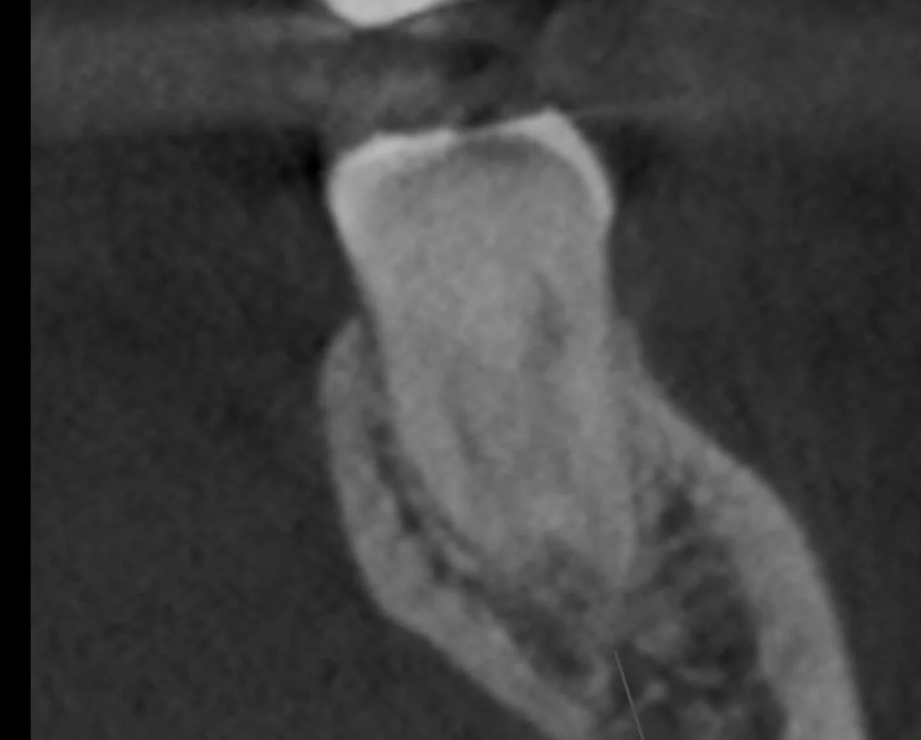
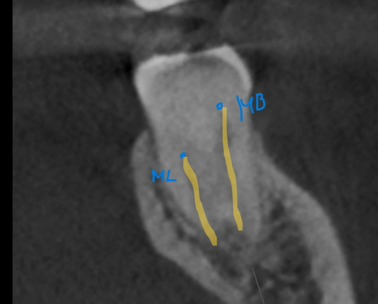
Coronal CBCT slice shows ML orifice likely to appears 3-4mm apical to floor of pulp chamber.
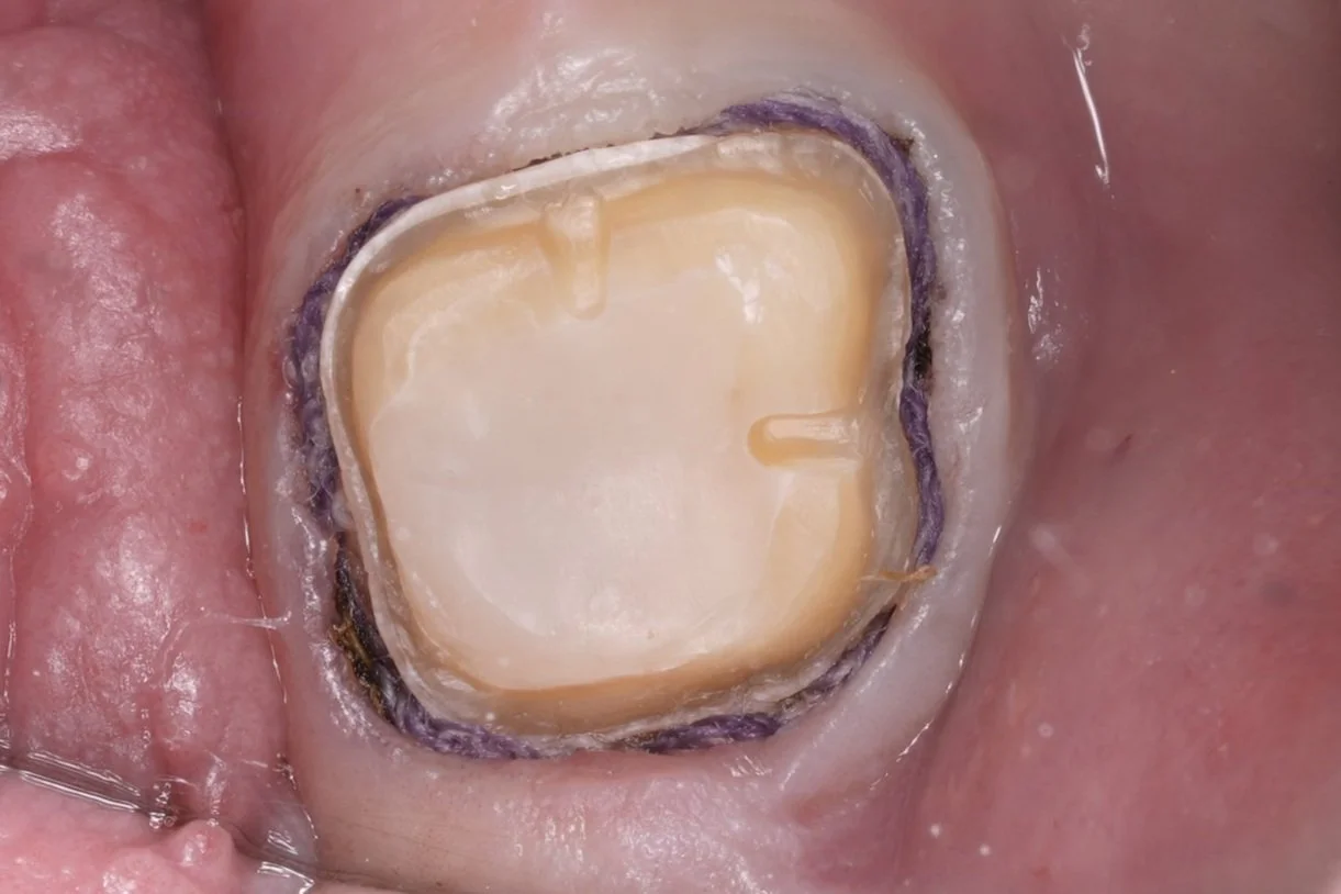
I needed to carefully expose the ML orifice using #10 pulp burs and used the pulp floor map as a guide.
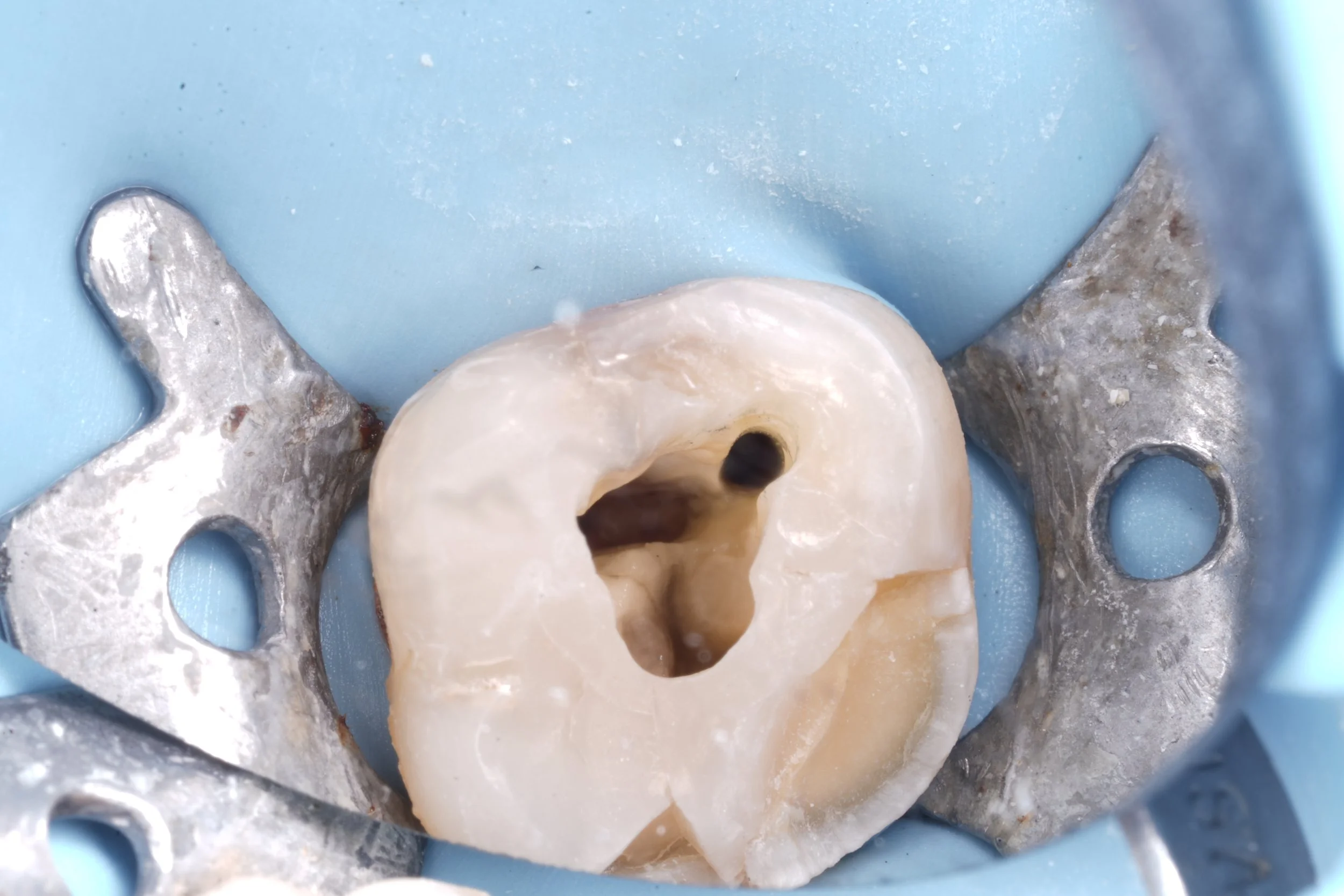

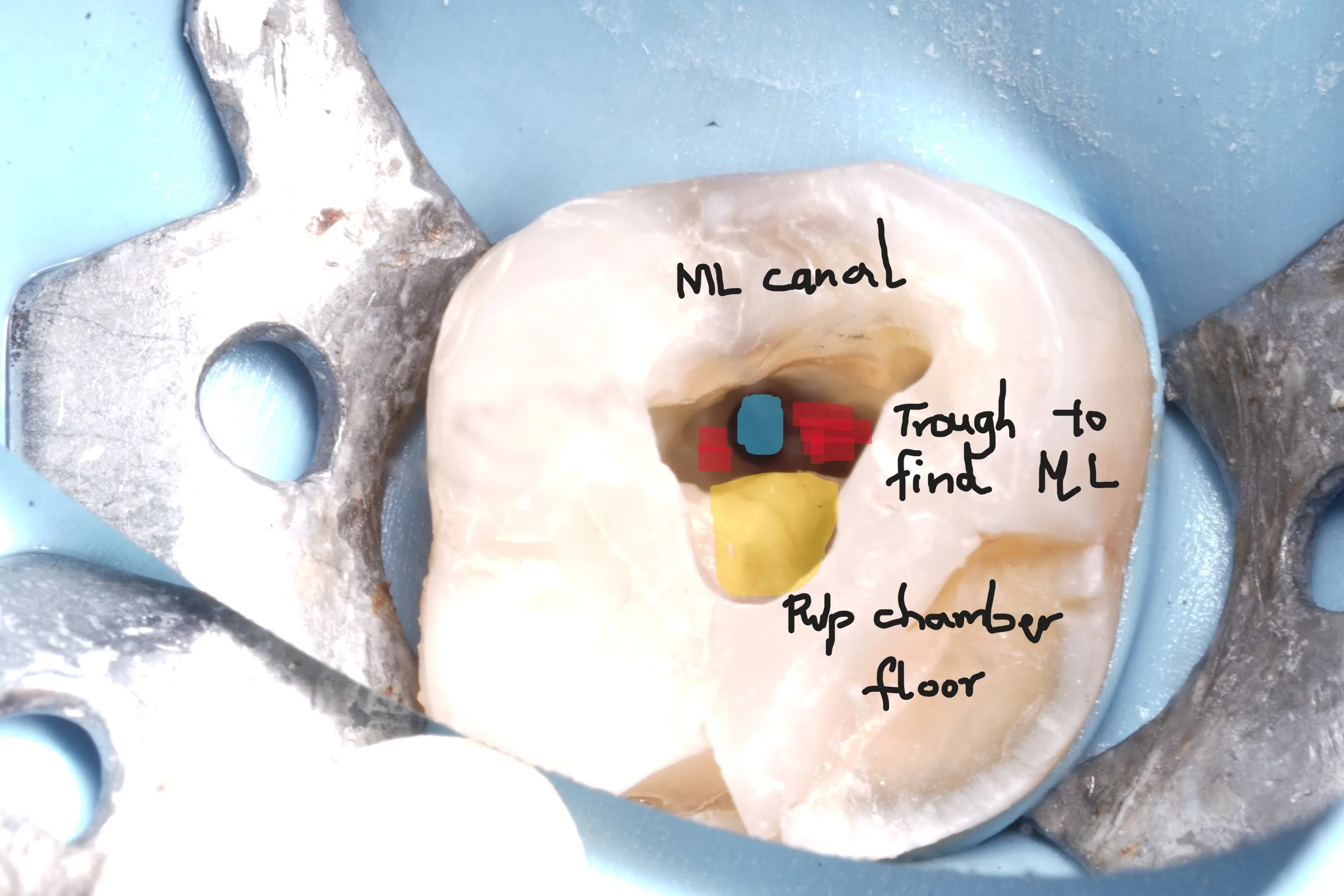
After troughing in the ML region, orifice was eventual exposed and canals prepared with rotary files.

Overall this was a difficult procedure and a large amount of time was needed for the visit. Searching for the ML was also challenging as the risk of perforation was ever present.